Showing posts with label Medical Research. Show all posts

4/27/2013

Tests on Hereditary Diseases

Scientists at the University of Medicine and Dentistry of New Jersey-New Jersey Medical School (UMDNJ-NJMS) have developed new DNA sequencing tests that hold significant promise for decreasing costs associated with diagnosing cancer and hereditary diseases, including cystic fibrosis.

According to the Cystic Fibrosis Foundation web site, "More than 10 million Americans are symptomless carriers of the defective CF gene." This chronic disease impacts the lungs and the digestive system. It occurs when a child inherits one defective CF gene from each parent. Statistics show New Jersey averages 125,000 births of children who are diagnosed with cystic fibrosis annually.Officials at the New Jersey Department of Health approved the use of the new Cystic Fibrosis (CF) Carrier and Diagnosis Test, which was created at the Institute for Genomic Medicine at UMDNJ-NJMS. Using a semiconductor mechanism that was developed by San Francisco-based Ion Torrent, the microchip tests the entire gene for mutations. IGM now offers this certified Clinical Diagnostic Laboratory service for hospitals as well as obstetrics and gynecology practices throughout the Garden State.
"We believe the adaptation of this new sequencing technology will drastically improve our ability to analyze genetic disorders," said Marvin N. Schwalb, PhD, director of the Institute for Genomic Medicine. "Traditional CF sequencing testing costs thousands of dollars making the test unavailable for carrier screening. This new test costs less than $200. Most importantly, the genetic carrier test we developed improves the diagnosis rate to 98 percent. While the test provides significant improvement for all populations, the improved rate is particularly valuable for minorities because current carrier screening methods only detects approximately 65% of mutations in these populations."
The new technology provides many advances including the ability to test as many as 96 samples on a single platform and the fact that the equipment cost 1/10 as much as the previous technology.
IGM has developed another test, which was also approved by the NJHSS, for mitochondrial DNA. Mutations in mitochondria cause a wide variety of diseases, such eye and neuromuscular system disorders and possible cancer.
Schwalb, a professor of Pediatrics, Microbiology and Molecular Genetics at NJMS, said, "We are proud of the fact that the IGM is a world leader in the advancement of genetic diagnosis. DNA sequencing will keep us very busy for a while. In the state of New Jersey, there is nothing that compares to this advancement and this is just the beginning."
Source: University of Medicine and Dentistry of New Jersey (UMDNJ) (2012, August 16). Researchers develop DNA sequencing tests for hereditary diseases. ScienceDaily. Retrieved April 27, 2013, from http://www.sciencedaily.com/releases/2012/08/120816170309.htm?utm_source=feedburner&utm_medium=feed&utm_campaign=Feed%3A+sciencedaily+%28ScienceDaily%3A+Latest+Science+News%29

4/25/2013

Gut bacteria linked to obesity

Researchers at the University of Maryland School of Medicine have identified 26 species of bacteria in the human gut microbiota that appear to be linked to obesity and related metabolic complications. These include insulin resistance, high blood sugar levels, increased blood pressure and high cholesterol, known collectively as "the metabolic syndrome," which significantly increases an individual’s risk of developing diabetes, cardiovascular disease and stroke.

"We identified 26 species of bacteria that were correlated with obesity and metabolic syndrome traits such as body mass index (BMI), triglycerides, cholesterol, glucose levels and C-reactive protein, a marker for inflammation," says the senior author, Claire M. Fraser, Ph.D., professor of medicine and microbiology and immunology and director of the Institute for Genome Sciences (IGS) at the University of Maryland School of Medicine. "We can’t infer cause and effect, but it’s an important step forward that we're starting to identify bacteria that are correlated with clinical parameters, which suggests that the gut microbiota could one day be targeted with medication, diet or lifestyle changes."The results of the study, which analyzed data from the Old Order Amish in Lancaster County, Pa., are being published online on Aug. 15, 2012, in PLOS ONE, which is published by the Public Library of Science (PLOS One). The study was funded by the National Institutes of Health (NIH). (UH2/UH3 DK083982, U01 GM074518 and P30 DK072488)
Dr. Fraser says that additional research, including an interventional study with the Amish, is essential. "We can look at whether these bacteria change over time in a given individual or in response to diet or medication," she says.
Dr. Fraser notes that the research team, led by Margaret L. Zupancic, Ph.D., then a postdoctoral fellow at IGS, also found an apparent link between the gut bacteria and inflammation, which is believed to be a factor in obesity and many other chronic diseases. "This is one of the first studies of obesity in humans to make a link between inflammatory processes and specific organisms that are present in the GI tract," Dr. Fraser says, noting that participants with metabolic syndrome who had elevated serum markers associated with inflammation tended to have the lowest levels of good bacteria that have been reported previously to have anti-inflammatory properties.
The study is the result of an ongoing collaboration between Dr. Fraser and Alan R. Shuldiner, M.D., in connection with the NIH’s Human Microbiome Project, which seeks to characterize microbial communities in the body. Dr. Shuldiner, associate dean for personalized medicine and director of the Program in Personalized and Genomic Medicine at the University of Maryland School of Medicine, operates an Amish research clinic in Lancaster Pa. Over the past 20 years, he and his research team have conducted more than a dozen studies with the Amish, looking for genes that may cause common diseases, such as diabetes, osteoporosis and cardiovascular disease.
"The Old Order Amish are ideal for such studies because they are a genetically homogenous population descended from a few founder families and have a similar rural lifestyle," Dr. Shuldiner, the John L. Whitehurst Professor of Medicine, says. "We believe the results of this study are relevant to a broader population because the clinical characteristics of obesity and its complications in the Amish are no different from the general Caucasian population," he says.
E. Albert Reece, M.D., Ph.D., M.B.A., vice president for medical affairs at the University of Maryland and the John Z. and Akiko K. Bowers Distinguished Professor and dean of the University of Maryland School of Medicine, says, "Obesity and its related complications have become a critical public health concern, and the number of people who are now considered obese or overweight has skyrocketed. Dr. Fraser and Dr. Shuldiner are two of our most senior research-scientists and leaders in their respective fields. This study provides valuable insights into the role the bacteria in our bodies may play in obesity and the metabolic syndrome. We may ultimately be able to target the gut microbiome to help prevent or mitigate risk factors for a number of diseases."
The researchers analyzed the bacteria in fecal samples of 310 members of the Old Order Amish community, using a process that enables them to identify a marker gene that serves as a bar code for each type of bacteria. Participants in the study ranged from lean to overweight to obese; some of the obese participants also had features of the metabolic syndrome. "Our hypothesis was that we would see a different composition in the gut microbiota in lean vs. obese individuals and possibly in individuals who were obese but also had features of the metabolic syndrome."
They discovered that every individual possessed one of three different communities of interacting bacteria, each characterized by a dominant bacterial genus. Neither BMI nor any metabolic syndrome trait was specifically associated with any of these communities. Instead, differing levels of 26 less abundant bacterial species present in all individuals appeared to be linked to obesity and certain features of the metabolic syndrome.
Interestingly, researchers also analyzed people's gut bacteria by their occupation and found that those who had regular contact with livestock, such as farmers and their wives, had bacterial communities dominated by Prevotella, a type of bacteria that is also abundant in the gut microbiota of cattle and sheep. "These findings suggest that environmental exposure may play a role in determining the composition of the gut microbiota in humans," Dr. Fraser says.

University of Maryland Medical Center (2012, August 15). Gut bacteria linked to obesity and metabolic syndrome identified. ScienceDaily. Retrieved April 26, 2013, from http://www.sciencedaily.com­/releases/2012/08/120815174902.htm?utm_source=feedburner&utm_medium=feed&utm_campaign=Feed%3A+sciencedaily+%28ScienceDaily%3A+Latest+Science+News%29

4/24/2013

Scientists Can Now Block Heroin, Morphine Addiction

In a major breakthrough, an international team of scientists has proven that addiction to morphine and heroin can be blocked, while at the same time increasing pain relief.


Laboratory studies have shown that the drug (+)-naloxone (pronounced: PLUS nal-OX-own) will selectively block the immune-addiction response.The team from the University of Adelaide and University of Colorado has discovered the key mechanism in the body's immune system that amplifies addiction to opioid drugs.
The results -- which could eventually lead to new co-formulated drugs that assist patients with severe pain, as well as helping heroin users to kick the habit -- will be published August 16 in the Journal of Neuroscience.
"Our studies have shown conclusively that we can block addiction via the immune system of the brain, without targeting the brain's wiring," says the lead author of the study, Dr Mark Hutchinson, ARC Research Fellow in the University of Adelaide's School of Medical Sciences.
"Both the central nervous system and the immune system play important roles in creating addiction, but our studies have shown we only need to block the immune response in the brain to prevent cravings for opioid drugs."
The team has focused its research efforts on the immune receptor known as Toll-Like receptor 4 (TLR4).
"Opioid drugs such as morphine and heroin bind to TLR4 in a similar way to the normal immune response to bacteria. The problem is that TLR4 then acts as an amplifier for addiction," Dr Hutchinson says.
"The drug (+)-naloxone automatically shuts down the addiction. It shuts down the need to take opioids, it cuts out behaviours associated with addiction, and the neurochemistry in the brain changes -- dopamine, which is the chemical important for providing that sense of 'reward' from the drug, is no longer produced."
Senior author Professor Linda Watkins, from the Center for Neuroscience at the University of Colorado Boulder, says: "This work fundamentally changes what we understand about opioids, reward and addiction. We've suspected for some years that TLR4 may be the key to blocking opioid addiction, but now we have the proof.
"The drug that we've used to block addiction, (+)-naloxone, is a non-opioid mirror image drug that was created by Dr Kenner Rice in the 1970s. We believe this will prove extremely useful as a co-formulated drug with morphine, so that patients who require relief for severe pain will not become addicted but still receive pain relief. This has the potential to lead to major advances in patient and palliative care," Professor Watkins says.
The researchers say clinical trials may be possible within the next 18 months.
This study has been funded by the National Institute on Drug Abuse (NIDA) in the United States and the Australian Research Council (ARC).
Source: University of Adelaide (2012, August 14). Scientists can now block heroin, morphine addiction. ScienceDaily. Retrieved April 24, 2013, from http://www.sciencedaily.com­/releases/2012/08/120814213246.htm

4/23/2013

An Artificial Retina With the Capacity to Restore Normal Vision

Two researchers at Weill Cornell Medical College have deciphered a mouse's retina's neural code and coupled this information to a novel prosthetic device to restore sight to blind mice. The researchers say they have also cracked the code for a monkey retina -- which is essentially identical to that of a human -- and hope to quickly design and test a device that blind humans can use.


The lead researcher, Dr. Sheila Nirenberg, a computational neuroscientist at Weill Cornell, envisions a day when the blind can choose to wear a visor, similar to the one used on the television show Star Trek. The visor's camera will take in light and use a computer chip to turn it into a code that the brain can translate into an image.The breakthrough, reported in theProceedings of the National Academy of Sciences (PNAS), signals a remarkable advance in longstanding efforts to restore vision. Current prosthetics provide blind users with spots and edges of light to help them navigate. This novel device provides the code to restore normal vision. The code is so accurate that it can allow facial features to be discerned and allow animals to track moving images.
"It's an exciting time. We can make blind mouse retinas see, and we're moving as fast as we can to do the same in humans," says Dr. Nirenberg, a professor in the Department of Physiology and Biophysics and in the Institute for Computational Biomedicine at Weill Cornell. The study's co-author is Dr. Chethan Pandarinath, who was a graduate student with Dr. Nirenberg and is currently a postdoctoral researcher at Stanford University.
This new approach provides hope for the 25 million people worldwide who suffer from blindness due to diseases of the retina. Because drug therapies help only a small fraction of this population, prosthetic devices are their best option for future sight. "This is the first prosthetic that has the potential to provide normal or near-normal vision because it incorporates the code," Dr. Nirenberg explains.
Discovering the Code
Normal vision occurs when light falls on photoreceptors in the surface of the retina. The retinal circuitry then processes the signals from the photoreceptors and converts them into a code of neural impulses. These impulses are then sent up to the brain by the retina's output cells, called ganglion cells. The brain understands this code of neural pulses and can translate it into meaningful images.
Blindness is often caused by diseases of the retina that kill the photoreceptors and destroy the associated circuitry, but typically, in these diseases, the retina's output cells are spared.
Current prosthetics generally work by driving these surviving cells. Electrodes are implanted into a blind patient's eye, and they stimulate the ganglion cells with current. But this only produces rough visual fields.
Many groups are working to improve performance by placing more stimulators into the patient's eye. The hope is that with more stimulators, more ganglion cells in the damaged tissue will be activated, and image quality will improve.
Other research teams are testing use of light-sensitive proteins as an alternate way to stimulate the cells. These proteins are introduced into the retina by gene therapy. Once in the eye, they can target many ganglion cells at once.
But Dr. Nirenberg points out that there's another critical factor. "Not only is it necessary to stimulate large numbers of cells, but they also have to be stimulated with the right code -- the code the retina normally uses to communicate with the brain."
This is what the authors discovered -- and what they incorporated into a novel prosthetic system.
Dr. Nirenberg reasoned that any pattern of light falling on to the retina had to be converted into a general code -- a set of equations -- that turns light patterns into patterns of electrical pulses. "People have been trying to find the code that does this for simple stimuli, but we knew it had to be generalizable, so that it could work for anything -- faces, landscapes, anything that a person sees," Dr. Nirenberg says.
Vision = Chip Plus Gene Therapy
In a eureka moment, while working on the code for a different reason, Dr. Nirenberg realized that what she was doing could be directly applied to a prosthetic. She and her student, Dr. Pandarinath, immediately went to work on it. They implemented the mathematical equations on a "chip" and combined it with a mini-projector. The chip, which she calls the "encoder" converts images that come into the eye into streams of electrical impulses, and the mini-projector then converts the electrical impulses into light impulses. These light pulses then drive the light-sensitive proteins, which have been put in the ganglion cells, to send the code on up to the brain.
The entire approach was tested on the mouse. The researchers built two prosthetic systems -- one with the code and one without. "Incorporating the code had a dramatic impact," Dr. Nirenberg says. "It jumped the system's performance up to near-normal levels -- that is, there was enough information in the system's output to reconstruct images of faces, animals -- basically anything we attempted."
In a rigorous series of experiments, the researchers found that the patterns produced by the blind retinas in mice closely matched those produced by normal mouse retinas.
"The reason this system works is two-fold," Dr. Nirenberg says. "The encoder -- the set of equations -- is able to mimic retinal transformations for a broad range of stimuli, including natural scenes, and thus produce normal patterns of electrical pulses, and the stimulator (the light sensitive protein) is able to send those pulses on up to the brain."
"What these findings show is that the critical ingredients for building a highly-effective retinal prosthetic -- the retina's code and a high resolution stimulating method -- are now, to a large extent, in place," reports Dr. Nirenberg.
Dr. Nirenberg says her retinal prosthetic will need to undergo human clinical trials, especially to test safety of the gene therapy component, which delivers the light-sensitive protein. But she anticipates it will be safe since similar gene therapy vectors have been successfully tested for other retinal diseases.
"This has all been thrilling," Dr. Nirenberg says. "I can't wait to get started on bringing this approach to patients."
The study was funded by grants from the National Institutes of Health and Cornell University's Institute for Computational Biomedicine.
Both Drs. Nirenberg and Pandarinath have a patent application for the prosthetic system filed through Cornell University.
Source: Weill Cornell Medical College (2012, August 14). An artificial retina with the capacity to restore normal vision. ScienceDaily. Retrieved April 23, 2013, from http://www.sciencedaily.com­/releases/2012/08/120814213326.htm

4/15/2013

Half of Inhaled Soot Particles from Diesel Exhaust, Fires Gets Stuck in the Lungs


The exhaust from diesel-fueled vehicles, wood fires and coal-driven power stations contains small particles of soot that flow out into the atmosphere. The soot is a scourge for the climate but also for human health. Now for the first time, researchers have studied in detail how diesel soot gets stuck in the lungs. The results show that more than half of all inhaled soot particles remain in the body. (Credit: © Imagenatural / Fotolia)

The figure is higher than for most other types of particles. For example "only" 20 per cent of another type of particle from wood smoke and other biomass combustion gets stuck in the lungs. One explanation is that diesel soot is made up of smaller particles and can therefore penetrate deeper into the lungs, where it is deposited. The study was made on diesel particles (which mainly consist of soot) and was recently published in the Journal of Aerosol Science. Ten healthy people volunteered for the the study.
"Findings of this kind can be extremely useful both for researchers to determine what doses of soot we get into our lungs out of the amount we are exposed to, and to enable public authorities to establish well-founded limits for soot particles in outdoor air," says Jenny Rissler, researcher in aerosol technology at Lund University's Faculty of Engineering and responsible for publishing the study.
In population studies, other researchers have been able to observe that people who live in areas with high concentrations of particulates are more affected by both respiratory and cardiovascular diseases. But since there is no conclusive evidence that it is precisely the soot that is to blame, the authorities have so far not taken any decisions on guidelines.
"Currently there is no specific limit for soot particles in the air, despite the fact that soot in the air is linked to both lung cancer and other diseases," says Jenny Rissler.
But Jenny Rissler thinks that in the future, limits on soot levels will also be set, with reference to the WHO's recent reclassification of diesel exhaust from "probably carcinogenic" to "carcinogenic."
Soot particles are not only connected to effects on health but may also contribute to a warmer climate. Paradoxically, other types of aerosol particles can partly be desirable, insofar as they have a cooling effect on the climate and thereby mitigate the warming effect of carbon dioxide.
"Soot particles are black and absorbs light, thus producing a warming effect. So it could be a double advantage to reduce it," she observes.
Jenny Rissler will next be studying individual variations in lung deposition and exposing cells to soot. She is also in the process of further developing methods to measure the surface area of the particles, as this has shown to be an important indicator of their harmfulness.
Background: Every time we breathe, we inhale tiny airborne particles, so-called aerosol particles. Some occur naturally, while others are the result of human activity. Soot mainly belongs in the latter category, as a by-product of combustion from power stations to small-scale wood fires and decorative candles. Another common source of soot is the exhaust from diesel engines, even though modern diesel cars have considerably reduced emissions thanks to efficient filters.
The EU will be tightening rules on emissions for heavy duty diesel vehicles in 2014.
-------------
The above story is reprinted from materials provided byLund University.
Jenny Rissler, Erik Swietlicki, Agneta Bengtsson, Christoffer Boman, Joakim Pagels, Thomas Sandström, Anders Blomberg, Jakob Löndahl. Experimental determination of deposition of diesel exhaust particles in the human respiratory tractJournal of Aerosol Science, 2012; 48: 18 DOI: 10.1016/j.jaerosci.2012.01.005
Lund University (2012, June 27). Half of inhaled soot particles from diesel exhaust, fires gets stuck in the lungs. ScienceDaily. Retrieved April 15, 2013, from http://www.sciencedaily.com­/releases/2012/06/120627092016.htm

4/11/2013

New research suggests animal-to-human transmission of MRSA

"Using whole genome sequencing, scientists have found two independent human cases of infection have been linked to livestock.

Having found this new MRSA in both people and animals on the same farm it was likely that it is being transmitted between animals and people.
Mark Holmes
Cambridge scientists have linked two human cases of infection with the antibiotic- resistant superbug MRSA to farms in Denmark. The results of the study, published today in the journal EMBO Molecular Medicine, suggest the methicillin-resistantStaphylococcus aureus (MRSA) bacteria was transmitted from the livestock to the farmers.
The type of MRSA which was found in both of the human cases was only discovered two years ago by Dr Mark Holmes and his colleagues from the University of Cambridge. The new strain’s genetic makeup differs greatly from previous strains, which means that the ‘gold standard’ molecular tests currently used to identify MRSA - a polymerase chain reaction technique (PCR) and slide agglutination testing - do not detect it.
For this study, the scientists used whole genome sequencing to investigate two cases of the new MRSA where the patients lived on farms to see if the same strain could be found in the animals on the farm.
Dr Holmes, from Cambridge’s Department of Veterinary Medicine and the senior author on the paper, said: “Having found this new MRSA in both people and animals on the same farm it was likely that it is being transmitted between animals and people.
“By looking at the single differences in nucleotides, or SNPs, in the DNA sequences of each isolate, it became obvious that in both farms we looked at the human and animal MRSA were almost identical. In one case, the results also clearly showed that the most likely direction of transmission was from animal to human.”
The study raises questions about whether cows could be a reservoir for new strains of MRSA. It was previously not clear whether MRSA was transmitted to cows from humans or to humans from cows, but the new research indicates that the livestock is the likely source of these new strains.
“Our findings demonstrate that the MRSA strains we studied are capable of transmission between animals and humans, which highlights the role of livestock as a potential reservoir of antibiotic resistant bacteria,” remarked Dr Ewan Harrison, a post-doctoral research associate at the University of Cambridge’s Department of Veterinary Medicine and co-author of the paper.
For more information about this story, please contact: Genevieve Maul, Office of Communications, University of Cambridge. Email: Genevieve.Maul@admin.cam.ac.uk; Tel: 01223 765542."

6/21/2012

Avian Flu Viruses Which Are Transmissible Between Humans Could Evolve in Nature


It might be possible for human-to-human airborne transmissible avian H5N1 influenza viruses to evolve in nature, new research has found.
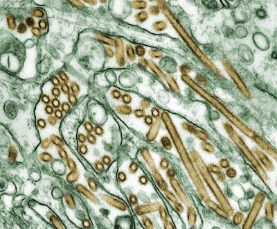
Avian Flu Viruses Which Are Transmissible Between Humans Could Evolve in Nature
Colorized transmission electron micrograph of Avian influenza A H5N1 viruses (seen in gold) grown in MDCK cells (seen in green) [Credit: CDC/Courtesy of Cynthia Goldsmith; Jacqueline Katz; Sherif R. Zaki]
The findings, from research led by Professor Derek Smith and Dr Colin Russell at the University of Cambridge, were published June 22 in the journal Science.

Currently, avian H5N1 influenza, also known as bird flu, can be transmitted from birds to humans, but not (or only very rarely) from human to human. However, two recent papers by Herfst, Fouchier and colleagues in Science and Imai, Kawaoka and colleagues in Nature reveal that potentially with as few as five mutations (amino acid substitutions), or four mutations plus reassortment, avian H5N1 can become airborne transmissible between mammals, and thus potentially among humans. However, until now, it was not known whether these mutations might evolve in nature.

The Cambridge researchers first analysed all of the surveillance data available on avian H5N1 influenza viruses from the last 15 years, focusing on birds and humans. They discovered that two of the five mutations seen in the experimental viruses (from the Fouchier and Kawaoka labs) had occurred in numerous existing avian flu strains. Additionally, they found that a number of the viruses had both of the mutations.

Colin Russell, Royal Society University Research Fellow at the University of Cambridge, said: "Viruses that have two of these mutations are already common in birds, meaning that there are viruses that might have to acquire only three additional mutations in a human to become airborne transmissible. The next key question is 'is three a lot, or a little?' "

The scientists explored this key question using a mathematical model of how viruses replicate and evolve within a mammalian host and assessed the influence of various factors on whether the remaining three mutations could evolve in a single host or in a short chain of transmission between hosts

The factors that increased the likelihood of mutations evolving are:

1. Random mutation. The replication mechanisms of influenza viruses don't make perfect copies. On average, every time an influenza virus replicates itself it makes approximately one mutation somewhere in the genome of each new virus. In each infected human there will be billions of viruses, and thus with many viruses replicating, multiple mutations can accumulate within a single host.

2. Positive selection. If some of the remaining mutations help the avian virus to adapt to mammals, then those mutations will make the viruses more fit and thus will be positively selected and preferentially accumulate.

3. Long infection. The longer someone is infected and producing new viruses, the more time there is for mutations to accumulate.

4. Functionally equivalent substitutions. The sets of substitutions identified by Fouchier and Kawaoka are unlikely to be the only combinations of substitutions capable of producing an aerosol transmissible virus. The probability of emergence increases with the number of combinations.

5. Diversity in the within-bird virus population. Given all of the mutations there are likely to be within a host due to random mutation, it is possible that the viruses from a bird that infect a human might have a mutation that would not be detected by routine surveillance. For example, if 100 virus particles from a bird infect a human and one of those particles had a key mutation, it would increase the probability of the mutation reaching high levels within a host even though routine sequencing would not detect it.

6. Transmission between mammals. If mammals are capable of transmitting viruses that have some but not all of the necessary substitutions it could increase the probability of an airborne transmissible virus evolving.

The factors that decreased the likelihood of mutations evolving are:

1. An effective immune response. An effective immune response would shorten the length of an infection and thus decrease the time available to accumulate mutations.

2. Deleterious substitutions. If any of the substitutions necessary for airborne transmission were harmful to the virus it would, on average, slow the accumulation of mutations.

3. Order of acquiring mutations. It is not currently known if the mutations for airborne transmissibility need to be acquired in a specific order. If they do, it would, on average, slow the accumulation of mutations.

"With the information we have, it is impossible to say what the exact risk is of the virus becoming airborne transmissible among humans. However, the results suggest that the remaining three mutations could evolve in a single human host, making a virus evolving in nature a potentially serious threat," said Derek Smith, Professor of Infectious Disease Informatics at the University of Cambridge. "We now know that it is in the realm of possibility that these viruses can evolve in nature, and what needs to be done to assess the risk more accurately of these mutations evolving in nature."

The scientists recommend the following activities be considered high priority for estimating and ameliorating the risk of emergence of aerosol transmissible H5N1 viruses.

First, additional surveillance in regions where viruses with airborne transmission enabling substitutions have been observed and in regions connected to those regions by bird migration and trade. Also, increased surveillance for mutations that might have the same function as those found by the Fouchier and Kawaoka labs.

Second, related to surveillance, some targeted sequencing of H5N1 viruses should be done by "deep sequencing" where the lab sequences many viruses from an individual host to look for viruses that might have accumulated the critical mutations, even if those viruses are just a small proportion of the viruses within an animal.

Third, further investigations are needed to determine which substitutions and combinations of substitutions that are not the same as, but have the same function as, the substitutions identified by the Fouchier and Kawaoka labs are capable of making viruses airborne transmissible between mammals.

Fourth, further studies are needed to elucidate the changes in within-host fitness and between-host transmissibility associated with each airborne transmission enabling substitution and combination of substitutions.

Professor Smith added: "The situation is similar to assessing the risk of an earthquake or tsunami. We don't know exactly when and where, but by increasing monitoring and research -- some of which is already underway -- scientists and public health officials will be able to increase the accuracy with which the risk can be assessed and to minimise those risks."

The research was funded by multiple sources including the European Commission through framework 7 grants EMPERIE and ANTIGONE, the Royal Society, the Human Frontiers Science Program, the Wellcome Trust, and the National Institutes of Health.

Source: University of Cambridge [June 21, 2012]

6/06/2012

1 Million Billion Billion Billion Billion Billion Billion: Number of Undiscovered Drugs


A new voyage into "chemical space" -- occupied not by stars and planets but substances that could become useful in everyday life -- has concluded that scientists have synthesized barely one tenth of 1 percent of the potential medicines that could be made. The report, in the journal ACS Chemical Neuroscience, estimates that the actual number of these so-called "small molecules" could be 1 novemdecillion (that's 1 with 60 zeroes), 1 million billion billion billion billion billion billion, which is more than some estimates of the number of stars in the universe.

1 Million Billion Billion Billion Billion Billion Billion: Number of Undiscovered Drugs
A new voyage into "chemical space" -- occupied not by stars and planets but substances that could become useful in everyday life -- has concluded that scientists have synthesized barely one tenth of 1 percent of the potential medicines that could be made [Credit: Web]
Jean-Louis Reymond and Mahendra Awale explain that small molecules, which are able to cross cell walls and interact with biological molecules in the body, are prime targets for scientists who develop new medicines. Most existing medications are small molecules. The authors focused on the "chemical space" inhabited by all of the small molecules that could possibly exist according to the laws of physics and chemistry. 

Researchers have identified millions of these compounds -- the ACS' Chemical Abstracts Service database contains almost 67 million substances. Reymond and Awale estimate that the molecules synthesized and tested as potential drugs so far represent less than 0.1 percent of chemical space. To aid researchers looking for new ways to prevent and treat disease, they set out to find the best ways to search for new small molecules.

The authors discuss several ways of getting a handle on chemical space, including by the size, shape and makeup of molecules. They show how computers can help researchers efficiently narrow a search for a new drug candidate. Computer modeling of chemical interactions can help researchers find a handful of promising molecules to synthesize and test in the lab. "Small molecule drugs are essential to the success of modern medicine," the authors note, and suggest that their methods may be particularly useful for finding new pharmaceuticals that target the central nervous system.

Source: American Chemical Society [June 06, 2012]

6/05/2012

Air Pollution Linked to Chronic Heart Disease


Air pollution, a serious danger to the environment, is also a major health risk, associated with respiratory infections, lung cancer and heart disease. Now a Tel Aviv University researcher has concluded that not only does air pollution impact cardiac events such as heart attack and stroke, but it also causes repeated episodes over the long term.

Air Pollution Linked to Chronic Heart Disease

Cardiac patients living in high pollution areas were found to be over 40 percent more likely to have a second heart attack when compared to patients living in low pollution areas, according to Dr. Yariv Gerber of TAU's School of Public Health at the Sackler Faculty of Medicine. "We know that like smoking cigarettes, pollution itself provokes the inflammatory system. If you are talking about long-term exposure and an inflammatory system that is irritated chronically, pollution may well be involved in the progression of atrial sclerosis that manifests in cardiac events," explains Dr. Gerber.

Done in collaboration with Prof. Yaacov Drory and funded by the Environmental and Health Fund in Jerusalem, the research was presented at the San Diego Epidemiological Meeting of the American Heart Association in March and the Annual Meeting of the Israeli Heart Society in April.

Risking recurrence

Air pollution has previously been acknowledged as a factor in heart attack risk, as well as other health risks. The goal of this study, says Dr. Gerber, was to quantify that association and determine the long-term effects of air pollution on myocardial infarction (MI) patients. Their study followed 1,120 first-time MI patients who had been admitted to one of eight hospitals in central Israel between 1992 and 1993, all of whom were under the age of 65 at the time of admittance. The patients were followed up until 2011, a period of 19 years.

Air quality was measured at 21 monitoring stations inareas where the patients lived, and analyzed by a group of researchers at the Technion in Haifa. After adjusting for other factors such as socio-economic status and disease severity, the researchers identified an association between pollution and negative clinical outcomes, including mortality and recurrent vascular events such as heart attack, stroke and heart failure.

Compared to patients who lived in areas with the lowest recorded levels of pollution, those in the most polluted environment were 43 percent more likely to have a second heart attack or suffer congestive heart failure and 46 percent more likely to suffer a stroke. The study also found that patients exposed to air pollution were 35 percent more likely to die in the almost 20 year period following their first heart attack than those who were exposed to lower levels of pollution.

According to Dr. Gerber, the true impact of air pollution might be even stronger than this study shows. "Our method of assessing exposure does have limitations. Because we are using data from monitoring stations, it's a crude estimate of exposure, which most likely leads to an underestimation of the association," he warns. He estimates that air pollution could have double the negative impact with more precise measurement.

Identifying vulnerable groups

The results of the study not only indicate a health benefit for a public policy that curtails air pollution caused by industrial emissions and second hand smoke, but also call for heightened awareness by clinicians. Doctors should be making their patients aware of the risks of remaining in high pollution areas, suggesting that they work to limit their exposure, Dr. Gerber suggests.

Another purpose of this study was to begin identifying populations that are vulnerable to MI and re-occurring MI. Establishing the connection between air pollution and long-term risk for patients with cardiovascular diseases was an important step towards that goal.

Source: American Friends of Tel Aviv University [June 05, 2012]

5/29/2012

16th-Century Korean Mummy Provides Clue to Hepatitis B Virus Genetic Code


The discovery of a mummified Korean child with relatively preserved organs enabled an Israeli-South Korean scientific team to conduct a genetic analysis on a liver biopsy which revealed a unique hepatitis B virus (HBV) genotype C2 sequence common in Southeast Asia.

16th-Century Korean Mummy Provides Clue to Hepatitis B Virus Genetic Code
The ancient Korean mummy of a child provides clues to the hepatitis B virus genome [Credit: Seoul National Univesity]
Additional analysis of the ancient HBV genomes may be used as a model to study the evolution of chronic hepatitis B and help understand the spread of the virus, possibly from Africa to East-Asia. It also may shed further light on the migratory pathway of hepatitis B in the Far East from China and Japan to Korea as well as to other regions in Asia and Australia where it is a major cause of cirrhosis and liver cancer.

The reconstruction of the ancient hepatitis B virus genetic code is the oldest full viral genome described in the scientific literature to date. It was reported in the May 21 edition of the scientific journal Hepathology by a research team from the Hebrew University of Jerusalem's Koret School of Veterinary Medicine, the Robert H. Smith Faculty of Agriculture, Food and Environment; the Hebrew University's Faculty of Medicine, the Hadassah Medical Center's Liver Unit; Dankook University and Seoul National University in South Korea.

Carbon 14 tests of the clothing of the mummy suggests that the boy lived around the 16th century during the Korean Joseon Dynasty. The viral DNA sequences recovered from the liver biopsy enabled the scientists to map the entire ancient hepatitis B viral genome.

Using modern-day molecular genetic techniques, the researchers compared the ancient DNA sequences with contemporary viral genomes disclosing distinct differences. The changes in the genetic code are believed to result from spontaneous mutations and possibly environmental pressures during the virus evolutionary process. Based on the observed mutations rates over time, the analysis suggests that the reconstructed mummy's hepatitis B virus DNA had its origin between 3,000 to 100,000 years ago.

The hepatitis B virus is transmitted through the contact with infected body fluids , i.e. from carrier mothers to their babies, through sexual contact and intravenous drug abuse. According to the World Health Organization, there are over 400 million carriers of the virus worldwide, predominantly in Africa, China and South Korea, where up to 15 percent of the population are cariers of the virus. In recent years, universal immunization of newborns against hepatitis B in Israel and in South Korea has lead to a massive decline in the incidence of infection.

The findings are the result of a collaborative effort between Dr. Gila Kahila Bar-Gal of the Hebrew University of Jerusalem's Koret School of Veterinary Medicine; Prof. Daniel Shouval of the Hadassah Medical Center's Liver Unit and Hebrew University; Dr. Myeung Ju Kim of Dankook University, Seok Ju Seon Memorial Museum; Dr. Dong Hoon Shin of Seoul National University, College of Medicine ; Prof Mark Spigelman of the Hebrew University's Dept. of Parasitology and Dr. Paul R. Grant of University College of London,Dept. of Virology.

Source: Hebrew University of Jerusalem [May 29, 2012]

5/24/2012

Nanoparticles Seen as Artificial Atoms


In the growth of crystals, do nanoparticles act as "artificial atoms" forming molecular-type building blocks that can assemble into complex structures? This is the contention of a major but controversial theory to explain nanocrystal growth. A study by researchers at the U.S. Department of Energy (DOE)'s Lawrence Berkeley National Laboratory (Berkeley Lab) may resolve the controversy and point the way to energy devices of the future.

Nanoparticles Seen as Artificial Atoms
These are sequential color TEM images showing the growth of Pt3Fe nanorods over time, displayed as minutes:seconds. At the far right, twisty nanoparticle chains straighten and stretch into nanorods [Credit:  Haimei Zheng]
Led by Haimei Zheng, a staff scientist in Berkeley Lab's Materials Sciences Division, the researchers used a combination of transmission electron microscopy and advanced liquid cell handling techniques to carry out real-time observations of the growth of nanorods from nanoparticles of platinum and iron. Their observations support the theory of nanoparticles acting like artificial atoms during crystal growth.

"We observed that as nanoparticles become attached they initially form winding polycrystalline chains," Zheng says. "These chains eventually align and attach end-to-end to form nanowires that straighten and stretch into single crystal nanorods with length-to-thickness ratios up to 40:1. This nanocrystal growth process, whereby nanoparticle chains as well as nanoparticles serve as the fundamental building blocks for nanorods, is both smart and efficient."

Zheng is the corresponding author of a paper describing this research in the journal Science. The paper is titled "Real-Time Imaging of Pt3Fe Nanorod Growth in Solution." Co-authors are Hong-Gang Liao, Likun Cui and Stephen Whitelam.

If the near limitless potential of nanotechnology is to even be approached, scientists will need a much better understanding of how nano-sized particles can assemble into hierarchical structures of ever-increasing organization and complexity. Such understanding comes from tracking nanoparticle growth trajectories and determining the forces that guide these trajectories.

Through the use of transmission electron microscopy and liquid observation cells, scientists at Berkeley Lab and elsewhere have made significant progress in observing nanoparticle growth trajectories, including the oriented attachment of nanoparticles -- the chemical phenomenon that starts the growth of nanocrystals in solution. However, these observations have typically been limited to the first few minutes of crystal growth. In their study, Zheng and her colleagues were able to extend the time of observation from minutes to hours.

"The key to studying the growth of colloidal nanocrystals with different shapes and architectures is to maintain the liquid in the viewing window long enough to allow complete reactions," Zheng says. "We dissolved molecular precursors of platinum and iron in an organic solvent and used capillary pressure to draw the growth solution into a silicon-nitride liquid cell that we sealed with epoxy. The sealing of the cell was especially important as it helped keep the liquid from turning viscous over time. Previously, we'd often see the liquids become viscous and this would prevent the nanoparticle interactions that drive crystal growth from taking place."

Zheng and her colleagues chose to study the growth of platinum iron nanorods because of the electrocatalytic material's promising potential for use in next generation energy conversion and storage devices. They were able to observe these nanoparticles assemble into nanorod crystals using powerful transmission electron microscopes at Berkeley Lab's National Center for Electron Microscopy, including TEAM 0.5 (Transmission Electron Aberration-corrected Microscope), which can produce images with half‑angstrom resolution -- less than the diameter of a single hydrogen atom.

"From what we observed only single nanoparticles exist at the beginning of crystal growth, but, as growth proceeds, small chains of nanoparticles become dominant until, ultimately, only long chains of nanoparticles can be seen," Zheng says. "Our observations provide a link between the world of single molecules and hierarchical nanostructures, paving the way for the rational design of nanomaterials with controlled properties."

Source: DOE/Lawrence Berkeley National Laboratory [May 24, 2012]

Drug Destroys Human Cancer Stem Cells but Not Healthy Ones


A team of scientists at McMaster University has discovered a drug, thioridazine, successfully kills cancer stem cells in the human while avoiding the toxic side-effects of conventional cancer treatments.

Drug Destroys Human Cancer Stem Cells but Not Healthy Ones

"The unusual aspect of our finding is the way this human-ready drug actually kills cancer stem cells; by changing them into cells that are non-cancerous," said Mick Bhatia, the principal investigator for the study and scientific director of McMaster's Stem Cell and Cancer Research Institute in the Michael G. DeGroote School of Medicine.

Unlike chemotherapy and radiation, thioridazine appears to have no effect on normal stem cells.

The research, published May 24 in the science journal Cell, holds the promise of a new strategy and discovery pipeline for the development of anticancer drugs in the treatment of various cancers. The research team has identified another dozen drugs that have good potential for the same response.

For 15 years, some researchers have believed stem cells are the source of many cancers. In 1997, Canadian researchers first identified cancer stem cells in certain types of leukemia. Cancer stem cells have since been identified in blood, breast, brain, lung, gastrointestinal, prostate and ovarian cancer.

To test more than a dozen different compounds, McMaster researchers pioneered a fully automated robotic system to identify several drugs, including thioridazine.

"Now we can test thousands of compounds, eventually defining a candidate drug that has little effect on normal stem cells but kills the cells that start the tumor," said Bhatia.

The next step is to test thioridazine in clinical trials, focusing on patients with acute myeloid leukemia whose disease has relapsed after chemotherapy. Bhatia wants to find out if the drug can put their cancer into remission, and by targeting the root of the cancer (cancer stem cells) prevent the cancer from coming back. Researchers at McMaster have already designed how these trials would be done.

Bhatia's team found thioridazine works through the dopamine receptor on the surface of the cancer cells in both leukemia and breast cancer patients. This means it may be possible to use it as a biomarker that would allow early detection and treatment of breast cancer and early signs of leukemia progression, he said.

The research team's next step is to investigate the effectiveness of the drug in other types of cancer. In addition, the team will explore several drugs identified along with thioridazine. In the future, thousands of other compounds will be analyzed with McMaster robotic stem cell screening system in partnership with collaborations that include academic groups as well as industry.

"The goal for all of the partners is the same -- to find unique drugs to change the way we tackle and treat cancer," he said.

The research was supported by grants from the Canadian Institute of Health Research (CIHR), the Canadian Cancer Society Research Institute (CCSRI) and the Ontario Ministry of Economic Development and Innovation (MEDI)'s Ontario Consortium of Regenerating inducing Therapeutics (OCRiT).

Source: McMaster University [May 24, 2012]

5/19/2012

Genetic Discovery Will Revolutionize Understanding Of Gene Expression


Over the past decade, research in the field of epigenetics has revealed that chemically modified bases are abundant components of the human genome and has forced us to abandon the notion we've had since high school genetics that DNA consists of only four bases.

Genetic Discovery Will Revolutionize Understanding Of Gene Expression
Over the past decade, research in the field of epigenetics has revealed that chemically modified bases are abundant components of the human genome and has forced us to abandon the notion we've had since high school genetics that DNA consists of only four bases. Now, researchers have made a discovery that once again forces us to rewrite our textbooks. This time, however, the findings pertain to RNA, which like DNA carries information about our genes and how they are expressed. The researchers have identified a novel base modification in RNA which they say will revolutionize our understanding of gene expression [Credit: © Attila Németh / Fotolia]
Now, researchers at Weill Cornell Medical College have made a discovery that once again forces us to rewrite our textbooks. This time, however, the findings pertain to RNA, which like DNA carries information about our genes and how they are expressed. The researchers have identified a novel base modification in RNA which they say will revolutionize our understanding of gene expression.

Their report, published in the journal Cell, shows that messenger RNA (mRNA), long thought to be a simple blueprint for protein production, is often chemically modified by addition of a methyl group to one of its bases, adenine. Although mRNA was thought to contain only four nucleobases, their discovery shows that a fifth base, N6-methyladenosine (m6A), pervades the transcriptome. The researchers found that up to 20 percent of human mRNA is routinely methylated. Over 5,000 different mRNA molecules contain m6A, which means that this modification is likely to have widespread effects on how genes are expressed.

"This finding rewrites fundamental concepts of the composition of mRNA because, for 50 years, no one thought mRNA contained internal modifications that control function," says the study's senior investigator, Dr. Samie R. Jaffrey, an associate professor of pharmacology at Weill Cornell Medical College.

"We know that DNA and proteins are routinely modified by chemical switches that have profound effects on their function in both health and disease. But biologists believed mRNA was simply an intermediate between DNA and protein," he says. "Now we know mRNA is much more complex, and defects in RNA methylation can lead to disease."

Indeed, as part of the study, the researchers demonstrated that the obesity risk gene, FTO (fat mass and obesity-associated), encodes an enzyme capable of reversing this modification, converting m6A residues in mRNA back to regular adenosine. Humans with FTO mutations have an overactive FTO enzyme, which results in low levels of m6A and causes abnormalities in food intake and metabolism that lead to obesity.

The researchers uncovered links between m6A and other diseases as well.

"We found that m6A is present in many mRNAs encoded by genes linked to human diseases, including cancer as well as several brain disorders, such as autism, Alzheimer's disease, and schizophrenia," says the study's lead investigator, Dr. Kate Meyer, a postdoctoral researcher in Dr. Jaffrey's laboratory. 

"Methylation in RNA is a reversible modification that appears to be a central step in a wide variety of biological pathways and physiological processes," she says.

The first time that m6A was detected in mRNA was in 1975, but at the time scientists were unsure whether this finding was a result of contamination by other RNA molecules, Dr. Jaffrey says. Over 90 percent of RNA is either transfer RNA (tRNA) or ribosomal RNA (rRNA), cellular workhorses that are routinely modified.

But Dr. Jaffrey says he has always been interested in the idea that mRNA may be modified - "DNA, proteins, other forms of RNA are modified, so why not mRNA?" he says - so he and investigators in his laboratory developed a technique to help them uncover methylation in mRNA taken from both mouse and human samples.

They used two different antibodies that recognize and bind to m6A in mRNA in order to selectively isolate the mRNAs that contain m6A. By subjecting these mRNAs to next-generation sequencing, they were able to identify the sequence of each individual mRNA they had isolated. Co-authors Dr. Christopher Mason and Dr. Olivier Elemento, assistant professors from the Department of Physiology and Biophysics and Computational Genomics in Computational Biomedicine at Weill Cornell Medical College, then developed computational algorithms to reveal the identity of each of these methylated mRNAs.

The Weill Cornell researchers don't know how the thousands of m6As they detected in humans work to control the function of mRNAs, but they do note that the m6As are located near "stop codons" in mRNA sequences. These areas signal the end of translation of the mRNA, suggesting that m6A might influence ribosomal function. "But we really don't know yet," says Dr. Mason, a co-lead investigator on the study. "It may allow other proteins to bind to mRNA, or subject these mRNAs to a whole new regulatory pathway. Our bioinformatics analyses are providing several hints about the possible impact of methylation on RNA function."

Indeed, in their study, the investigators have already found that m6A sites frequently occur in regions of mRNA that are highly conserved across several species of vertebrates. "This shows that m6A sites are not just important for humans, but rather are maintained under selection across hundreds of millions of years of evolution, and thus are likely of critical importance for all animals," Dr. Mason says.

"This is the first demonstration of an epitranscriptomic modification - alterations in RNA function that are not due to changes in the underlying sequence," he adds.

"These findings are very, very exciting, and amazing, really, when you consider that mRNA has been around for so long and that nobody realized, in all this time, that they were being regulated in this way," Dr. Jaffrey says. "It was right under our noses."

In addition to investigating how m6A regulates mRNAs within cells, the researchers are now focused on identifying the enzymes and pathways that control mRNA methylation.

Their study already demonstrates that FTO is capable of reversing adenosine methylation and suggests that it acts on a large proportion of cellular mRNA. "FTO mutations are estimated to occur in one billion people worldwide and are a leading cause of obesity and type 2 diabetes. Our studies link m6A levels in mRNA to these major health problems and identify for the first time the mRNAs which are potentially targeted by FTO," Dr. Meyer says.

The investigators are currently working to understand how defective regulation of m6A in patients with FTO mutations causes obesity and metabolic disorders, and they are also developing tests to rapidly identify compounds that inhibit FTO activity. These compounds are expected to inhibit the overactive FTO found in humans, potentially leading to novel therapeutics for diabetes and obesity.

Source: Medical News Today {May 19, 2012]

5/16/2012

In the Genes, but Which Ones? Studies That Linked Specific Genes to Intelligence Were Largely Wrong, Experts Say


For decades, scientists have understood that there is a genetic component to intelligence, but a new Harvard study has found both that most of the genes thought to be linked to the trait are probably not in fact related to it, and identifying intelligence's specific genetic roots may still be a long way off.

In the Genes, but Which Ones? Studies That Linked Specific Genes to Intelligence Were Largely Wrong, Experts Say
For decades, scientists have understood that there is a genetic component to intelligence, but a new study has found both that most of the genes thought to be linked to the trait are probably not in fact related to it, and identifying intelligence's specific genetic roots may still be a long way off [Credit: Web]
Led by David I. Laibson '88, the Robert I. Goldman Professor of Economics, and Christopher F. Chabris '88, Ph.D. '99, assistant professor of psychology at Union College in Schenectady, N.Y., a team of researchers examined a dozen genes using large data sets that included both intelligence testing and genetic data. As reported in a forthcoming article in the journal Psychological Science, they found that in nearly every case, the hypothesized genetic pathway failed to replicate. In other words, intelligence could not be linked to the specific genes that were tested.

"It is only in the past 10 or 15 years that we have had the technology for people to do studies that involved picking a particular genetic variant and investigating whether people who score higher on intelligence tests tend to have that genetic variant," said Chabris. "In all of our tests we only found one gene that appeared to be associated with intelligence, and it was a very small effect. This does not mean intelligence does not have a genetic component, it means it's a lot harder to find the particular genes, or the particular genetic variants, that influence the differences in intelligence."

To get at the question of how genes influence intelligence, researchers first needed data, and plenty of it.

Though it had long been understood, based on studies of twins, that intelligence was a heritable trait, it wasn't until relatively recently that the technology emerged to allow scientists to directly probe DNA in a search for genes that affected intelligence.

The problem, Chabris said, was that early technology for assaying genes was very expensive, meaning that such studies were typically limited to, at most, several hundred subjects, who would take IQ tests and provide DNA samples for testing.

As part of their study, Chabris and his colleagues relied on several pre-existing data sets -- a massive study of Wisconsin high school graduates that began in the 1950s, the Framingham Heart Study, and an ongoing survey of all twins born in Sweden -- to expand that subject pool from a few hundred to many thousands.

"What we want to emphasize is that we are not saying the people who did earlier research in this area were foolish or wrong," Chabris said. "They were using the best technology they had available. At the time it was believed that individual genes would have a much larger effect -- they were expecting to find genes that might each account for several IQ points."

To identify genes that might play a role in intelligence, previous researchers used the "candidate gene approach," which requires identifying a gene that is already linked with a known biological function -- such as Alzheimer's disease or the production of a specific neurotransmitter. If people who scored high on intelligence tests shared a particular variant of that gene, it was believed, that demonstrated the gene's role in intelligence.

"These were reasonable hypotheses," said study co-author Daniel J. Benjamin '99, Ph.D. '06, assistant professor of economics at Cornell University. "But in retrospect, either the findings were false positives or the effects of the genes are much, much smaller than anyone had anticipated."

Chabris, however, emphasized that the results don't point to the idea that the dozen genes examined in the study play no role in intelligence, but rather suggest that intelligence may be tied to many genes and the ways in which they interact.

"As is the case with other traits, like height, there are probably thousands of genes and their variants that are associated with intelligence," he said. "And there may be other genetic effects beyond the single gene effects -- there could be interactions between genes, there could be interactions between genes and the environment. What our results show is that the way researchers have been looking for genes that may be related to intelligence -- the candidate gene method -- is fairly likely to result in false positives, so other methods should be used."

Author: Peter Reuell | Source: Harvard University [February 24, 2012]

5/14/2012

Powerful Function of Single Protein That Controls Neurotransmission Discovered


Scientists at Weill Cornell Medical College have discovered that the single protein -- alpha 2 delta -- exerts a spigot-like function, controlling the volume of neurotransmitters and other chemicals that flow between the synapses of brain neurons. The study, published online in Nature, shows how brain cells talk to each other through these signals, relaying thoughts, feelings and action, and this powerful molecule plays a crucial role in regulating effective communication.


In the study, the investigators also suggest how the widely used pain drug Lyrica might work. The alpha 2 delta protein is the target of this drug and the new work suggests an approach to how other drugs could be developed that effectively twist particular neurotransmitter spigots on and off to treat neurological disorders. The research findings surprised the research team, which includes scientists from University College London.

"We are amazed that any single protein has such power," says the study's lead investigator Dr. Timothy A. Ryan, professor of Biochemistry and associate professor of Biochemistry in Anesthesiology at Weill Cornell Medical College. "It is indeed rare to identify a biological molecule's function that is so potent, that seems to be controlling the effectiveness of neurotransmission."

The researchers found that alpha 2 delta determines how many calcium channels will be present at the synaptic junction between neurons. The transmission of chemical signals is triggered at the synapse by the entry of calcium into these channels, so the volume and speed of neurotransmission depends on the availability of these channels.

Researchers discovered that taking away alpha 2 delta from brain cells prevented calcium channels from getting to the synapse. "But if you add more alpha 2 delta, you can triple the number of channels at synapses," Dr. Ryan says. "This change in abundance was tightly linked to how well synapses carry out their function, which is to release neurotransmitters."

Before this study, it was known that Lyrica, which is used for neuropathic pain, seizures and fibromyalgia, binds to alpha 2 delta, but little was understood about how this protein works to control synapses.

Lifting up the Hood

Dr. Ryan is building what he calls a "shop manual" of neurological function, much of which centers on synaptic neurotransmission. In 2007 and 2008, he discovered crucial clues to how neurons repackage the chemicals used to signal across synapses. In 2011, Dr. Ryan discovered that distinct neurons differently tune the speed by which they package these chemicals. And in a recent study published April 29 in Nature Neuroscience, he described, for the first time, the molecular mechanisms at the synapse that control the release of dopamine, a crucial neurotransmitter.

"We are looking under the hood of these machines for the first time," he says. "Many neurological diseases are considered to arise from pathologies of synaptic function. The synapse is so complex; at least a few thousand genes control how they work. Repairing them through treatment requires that we understand how they work."

Dr. Ryan and his team often use two tools to conduct these studies -- they pin fluorescent tags on to molecules involved in synaptic function, and use ultra sensitive microscopy technology to watch these molecules up close and in real-time.

The researchers used the same toolkit to examine the function of calcium channels, which triggers neurotransmission. "At all synapses, the secretion of a neurotransmitter is driven by the arrival of an electric impulse, initiated by another neuron," Dr. Ryan says. When this impulse arrives at the nerve terminal it triggers the opening of calcium channels. The calcium that rushes in is the key trigger that drives a synapse to secrete its neurotransmitter.

"We have known for the past half century that calcium is a key controller of neurotransmission," he says. "Any small change in calcium influx has a big impact on neurotransmission."

Protein Acts like a Shipping Label

But the number of calcium channels at the synapse is not static. Neurons constantly replace worn out channels, and to do this, they build the channels in the neuron's cell body and then package them up and ship them to the nerve terminal. In some cases, that is a very long journey -- as much as a few feet, such as the distance between the brain and the base of the spinal cord or the length of a leg.

In the study, researchers tagged fluorescent proteins onto a gene that encodes protein that makes a calcium channel and delivered it to neurons. They then watched the progress of the newly formed channels as they made their way, from day four to day seven, from the bodies of neurons to the synapse.

They also manipulated the levels of alpha 2 delta, a suspected calcium channel partner, and discovered that when the protein was increased, more calcium channels were moved to the synapse. Less alpha 2 delta reduced the flow. "We discovered that alpha 2 delta made the decision of how many calcium channels should be shipped the length of the neuron to the synapse," Dr. Ryan says. "It's like the channels couldn't be transported without an alpha 2 delta shipping label."

The research team found however that alpha 2 delta must work in at least two steps. When they impaired a piece of alpha 2 delta that resembles proteins that are involved in how cells bind to each other, they found that this broken alpha 2 delta could still help get calcium channels shipped down to synapses. But once there, they no longer helped drive neurotransmitter release. "This means that not only does alpha 2 delta help to get calcium channels shipped out, but it also implies that something at the synapse has to sign-off on receiving the calcium channels, putting them in the right place for them to do their job," Dr. Ryan says.

The researchers suggest that Lyrica might work by interfering with this final step since the piece of alpha 2 delta they "broke" that prevents the signing-off resembles parts of proteins that allows them to stick to each other in a kind of handshake.

These findings suggest that future therapies designed to manipulate neurotransmission could try to target this handshaking process, Dr. Ryan says. To do this will require that researchers identify the missing partner in the handshake.

"We hope these exciting findings are providing a new direction in how to make better drugs to control communication between brain cells," Dr. Ryan says.

The study was funded by the National Institutes of Mental Health and the Welcome Trust. Co-authors of the study include Dr. Michael B. Hoppa from Weill Cornell Medical College, and Dr. Beatrice Lana, Dr. Wojciech Margas, and Dr. Annette C. Dolphin from University College London.

Source: NewYork-Presbyterian Hospital/Weill Cornell Medical Center/Weill Cornell Medical College [May 13, 2012]

Twitter Delicious Facebook Digg Stumbleupon Favorites More

 
Design by Free WordPress Themes | Bloggerized by Lasantha - Premium Blogger Themes | Facebook Themes